(M.K. Miller and G.D.W. Smith, ATOM PROBE MICROANALYSIS, Materials Research Society, 1989.)
The microscope consists essentially of a vacuum vessel, across one end of which is located a phosphor screen. The specimen of the material to be studied is prepared into a needle-like form, with an end radius typically 50 - 100 nm, and is mounted along the axis of the vakuum vessel about 50 mm from the screen. The specimen is kept at cryogenic temperatures and is mounted on an electrical insulator so that it can be raised to a high positive potential (3 - 30 kV). A small amount of an inert gas, usually helium or neon, is admitted to the microscope vessel. When the potential on the specimen is increased, gas atoms in the vicinity of the specimen apex become polarized by the strong field and are drawn towards the surface.
If the field is sufficiently high (a few tens of volts per nanometer), the first gas atoms to reach the surface become field adsorbed in high-field sites above the apices of prominent surface atoms. Gas atoms which arrive subsequently will migrate across the surface above the field adsorbed layer until they are ionized by the quantum-mechanical tunneling process of field ionisation. In this process, an electron from the gas atom tunnels through the surface potential barrier into a vacant energie level in the specimen, leaving a positively charged gas ion above the surface. Such ions are then repelled from the sample towards the phosphor screen where they built up a highly magnified, projected image of the surface at which they were formed. The process of field ionisation occurs most readily above the more prominent surface atoms, and narrow beams of ions formed above such single atoms give rise to individual image spots on the phosphor screen.
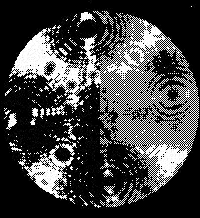
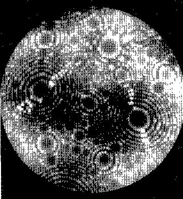
Examples of field ion micrographs obtained using microcannel plate image intensification at 15 kV in Helium at 78 K: (a) tungsten; (111) orientation and (b) tungsten; (100) orientation.
Members of the Interferometrygroup